
When Is the Best Time for a 4D Baby Scan During Pregnancy?
Watching a tiny human move inside the womb remains one of the most remarkable milestones of parenthood. While traditional two-dimensional scans offer critical medical data, a live 4D baby ultrasound Perth session adds a vivid dimension of depth, capturing real-time motions like yawning, stretching, and smiling.
Timing plays a crucial role in the outcome of these images. Scanning too early can reveal limited facial structure, while waiting too late might mean the baby becomes tightly wedged against the uterine wall. Finding the ideal window ensures clear visual detail and an unforgettable bonding experience.
Understanding the Difference Between 2D, 3D, and 4D Ultrasound
Ultrasound technology has evolved rapidly over recent decades. Each modality serves a unique purpose during the gestational journey.
Two-Dimensional (2D) Scans
Standard medical scans rely on 2D imaging. These grey, cross-sectional outlines allow sonographers to evaluate internal structures, measure organs, and assess fluid levels. They form the foundation of clinical prenatal care.
Three-Dimensional (3D) Scans

3D imaging takes multiple 2D cross-sections and compiles them into a stationary, three-dimensional photo. This technology displays external surface contours, showing the distinct shape of a nose, lips, or chubby cheeks.
Four-Dimensional (4D) Scans
Four-dimensional imaging introduces time as the fourth element. A 4D baby scan Perth session streams live 3D images, allowing parents to view continuous motion in real time.
The Ideal Window for a 4D Scan
Timing dictates the visual quality of three-dimensional and four-dimensional imaging. 4D scan Perth development, fetal positioning, and amniotic fluid volume all change dramatically across pregnancy stages.
Early Stage: Weeks 14 to 22
During this initial period, embryonic structures solidify into recognisable human features.
Facial features remain delicate as subcutaneous fat has not fully accumulated.
Fluid levels are high, offering ample space for the baby to move around freely.
Full-body views are easier to capture in a single frame.
Gender determination is highly reliable during these mid-trimester weeks.
The Peak Period: Weeks 26 to 30
This window represents the absolute sweet spot for detailed facial rendering.
Fat deposits under the skin increase, creating full cheeks and smooth facial contours.
Ample amniotic fluid surrounds the baby, creating the clear visual pathway required for sound waves.
Spatial room remains adequate before the baby settles deeply into the pelvis.
Subtle expressions such as sucking a thumb or blinking become clearly visible.
Late Stage: Weeks 31 to 34
Scanning remains possible during these later weeks, though visual clarity depends heavily on baby position.
Facial features display full newborn definition and volume.
Available space decreases as growth accelerates rapidly toward full term.
Reduced fluid margins around the face make clear captures more dependent on optimal positioning.
Key Factors That Impact Image Clarity
Clear imaging requires more than just scheduling the appointment at the right week. Several natural biological factors influence soundwave reflection and image crispness.
1. Amniotic Fluid Levels
Amniotic fluid acts as a transparent window for ultrasound waves. Clear space in front of the facial area produces crisp rendering. Reduced fluid pockets make features appear blurred against surrounding tissue.
2. Placental Location
An anterior placenta rests along the front wall of the uterus. While completely normal, this placement can sometimes sit close to the facial surface, requiring specialised angles to capture clear features.
3. Baby Position
Movement remains entirely up to the baby during the appointment. A face-down or rear-facing posture makes facial captures challenging. A gentle walk or a small sip of cold water can encourage a subtle shift in position.
4. Maternal Tissue Thickness
Ultrasound waves travel through abdominal tissue layers before reaching the uterus. Tissue density alters how sound waves travel, which can influence overall image sharpness.
How to Prepare for Your Appointment
Proper preparation optimises the chances of getting clear, high-definition images during your visit.
Maintain Consistent Hydration
Drink plenty of water for four to five days leading up to the session. Proper hydration improves amniotic fluid clarity, creating a clearer path for sound waves.
Eat a Balanced Light Snack
Consuming a small snack about twenty minutes before your scan can gently elevate blood sugar and encourage subtle movement.
Wear Comfortable Clothing
Opt for a two-piece outfit such as a top and trousers. This setup allows easy access to the abdomen while keeping you comfortable throughout the session.
Arrive Relaxed
Stress can affect muscle tension. Arriving calm helps foster a comfortable environment for both parent and baby.
Benefits of Choosing a 4D Ultrasound Experience
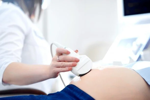
While diagnostic scans focus strictly on clinical health checks, elective four-dimensional imaging prioritises connection and family participation.
Enhanced Emotional Bonding: Seeing realistic facial features and live movements builds a deep, early emotional connection before birth.
Family Involvement: Spouses, partners, and extended family members can share in a relaxed, non-clinical environment.
Keepsake Memories: High-definition video clips and digital images create lasting records of early development.
Reassurance for Parents: Watching natural movements like stretching or resting brings peace of mind to expecting families.
Conclusion
Choosing the right time for a 4D scan Perth experience transforms an ordinary appointment into an unforgettable milestone. While early sessions offer full-body views, the window between 26 and 30 weeks provides the ideal balance of facial detail, fluid space, and active movement. Proper hydration, thoughtful preparation, and realistic expectations ensure a rewarding viewing experience. For parents in Western Australia seeking clear, high-quality bonding sessions, Pregnancy Ultrasound Perth offers a calm, welcoming environment to meet your little one before arrival.
FAQs
Why are weeks 26 to 30 considered the best time for a 4D baby scan?
During weeks 26 to 30, the baby has accumulated enough subcutaneous fat under the skin to develop full cheeks and smooth facial features. Additionally, there is still ample amniotic fluid surrounding the baby and sufficient space before they settle into the pelvis, creating the ideal conditions for clear, detailed 4D imaging and facial expressions.
Can I still get a clear 4D scan after 30 weeks of pregnancy?
Yes, scanning is still possible between weeks 31 and 34, and the baby’s face will display full newborn definition. However, because space in the womb decreases and fluid levels drop in later weeks, getting a clear image depends heavily on whether the baby is in an optimal position away from the uterine wall or placenta.
How does drinking extra water before the appointment help improve image quality?
Drinking plenty of water for 4 to 5 days leading up to your session increases the volume and clarity of your amniotic fluid. Amniotic fluid acts as a transparent window for ultrasound waves; clearer fluid surrounding the baby's face allows sound waves to reflect properly, resulting in sharper 3D and 4D imagery.
What is the main difference between a clinical 2D scan and an elective 4D scan?
A 2D scan produces standard cross-sectional, black-and-white outlines used by medical professionals to check internal structures, growth, and fetal health. An elective 4D scan streams live 3D video in real time, focusing on visual bonding, facial detail, and live movements like yawning or smiling in a relaxed, non-clinical setting.